Parts Parts And Functions Of A Microscope
The web page titled "Parts of a Microscope with Labeled Diagram and Functions" has the following key takeaways: 🔍 The microscope is an essential tool for scientists, researchers, and medical professionals. 🧬 The main function of a microscope is to provide a magnified view of small objects or organisms, such as bacteria, cells, or tissues.
1.5 Microscopy Biology LibreTexts
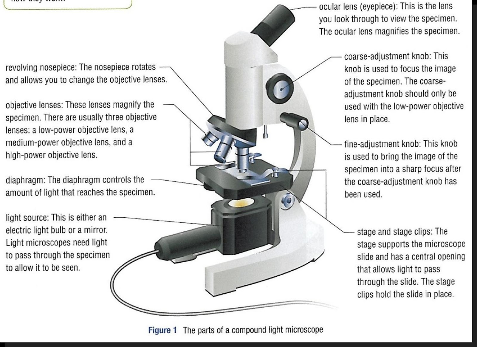
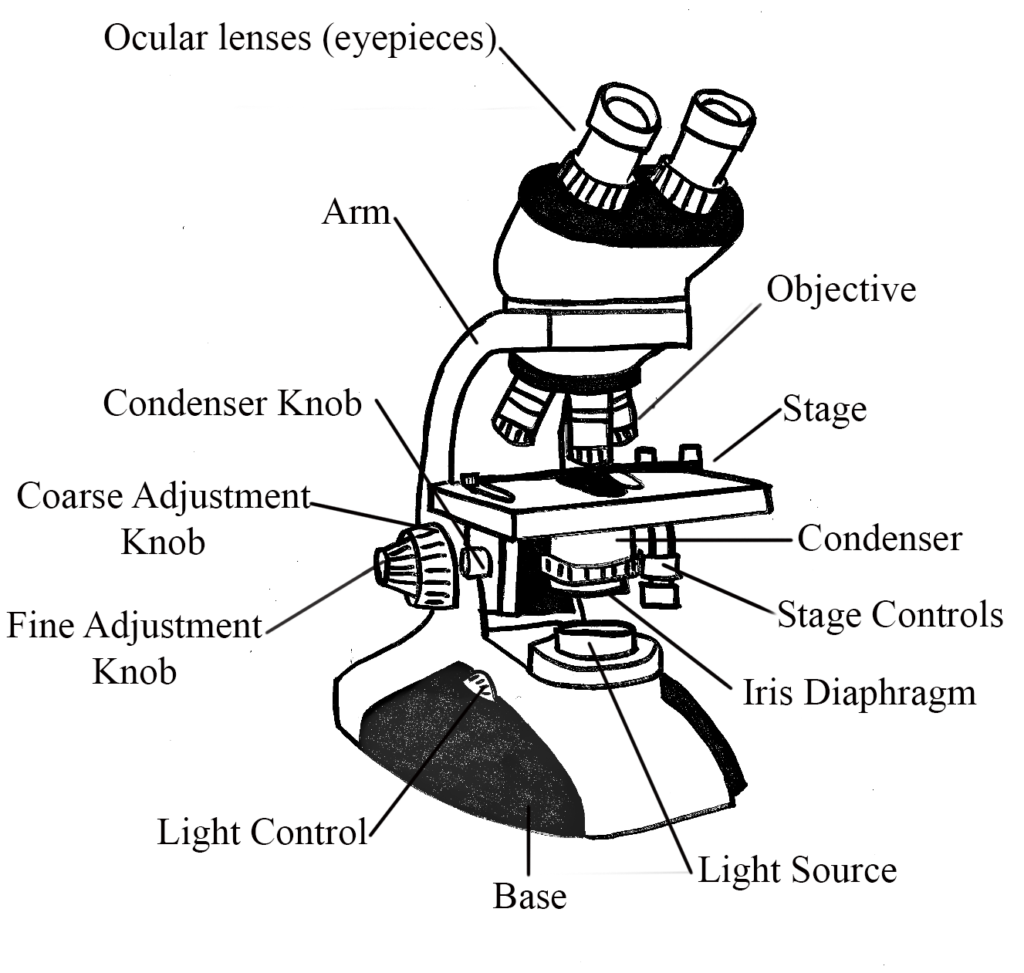
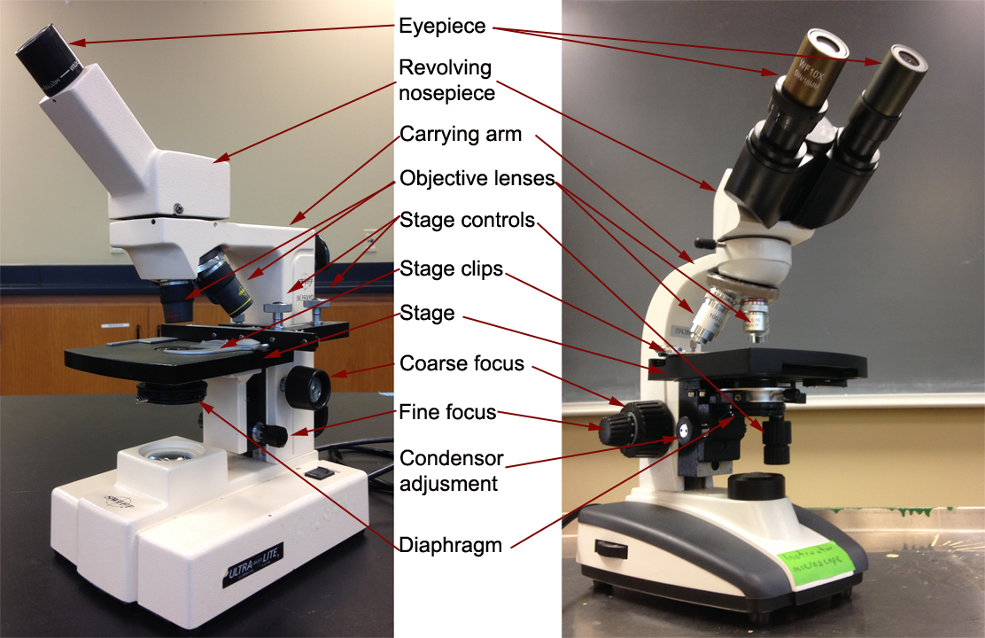
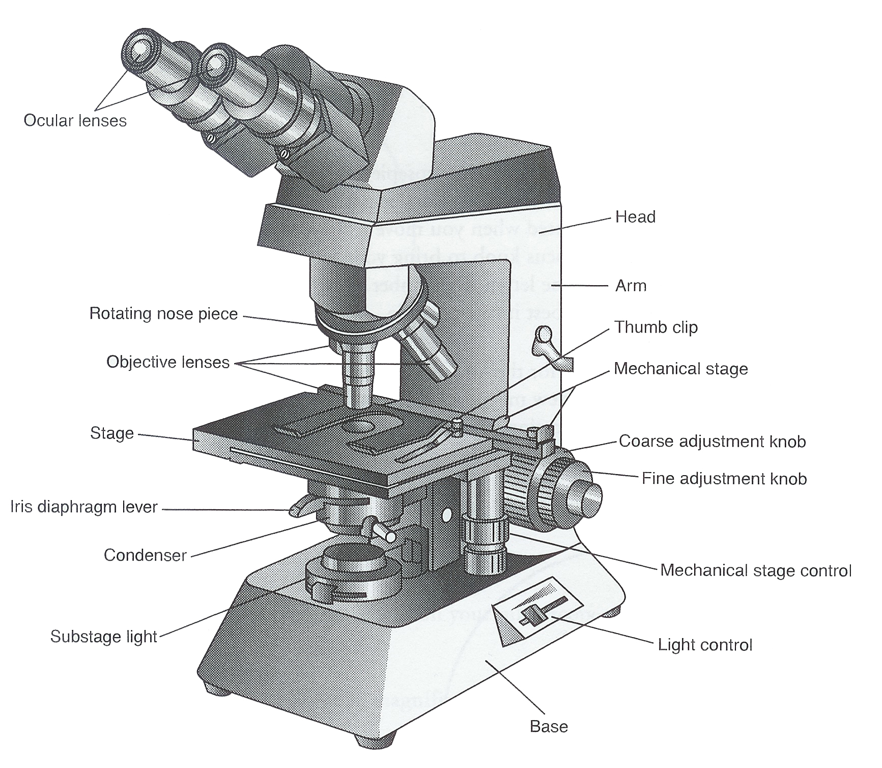
Figure: Diagram of parts of a microscope. There are three structural parts of the microscope i.e. head, arm, and base. Head - The head is a cylindrical metallic tube that holds the eyepiece lens at one end and connects to the nose piece at other end. It is also called a body tube or eyepiece tube.
Microscopes General Microbiology
Explore the different parts of a microscope using a diagram, including the microscope lens, eyepiece, and stage. Updated: 10/13/2022 Create an account
The Parts of a Compound Microscope and How To Handle Them Correctly
Simple Microscope Diagram (Parts) with Labels Frequently Asked Questions Definition and Working principle Simple microscope is a magnification apparatus that uses a combination of double convex lens to form an enlarged, erect image of a specimen.
Parts of a Compound Microscope — Learning in Hand with Tony Vincent
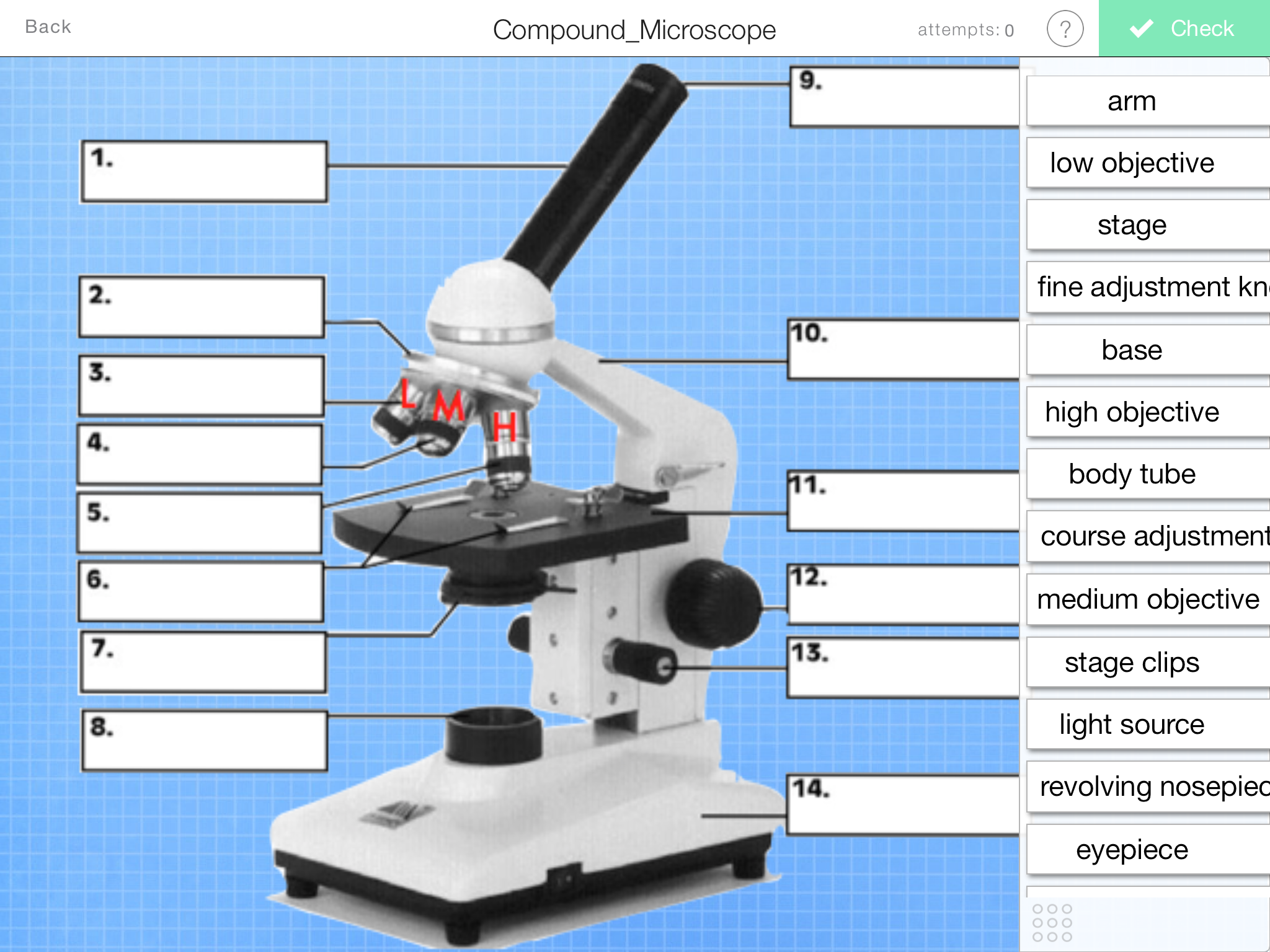
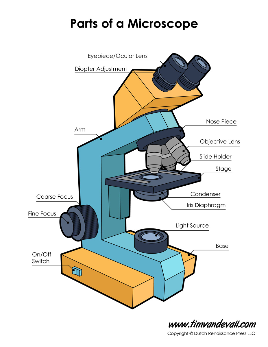
Label the microscope Interactive Add to collection Use this interactive to identify and label the main parts of a microscope. Drag and drop the text labels onto the microscope diagram. base high-power objective eye piece lens coarse focus adjustment diaphragm or iris stage fine focus adjustment light source Download Exercise Tweet
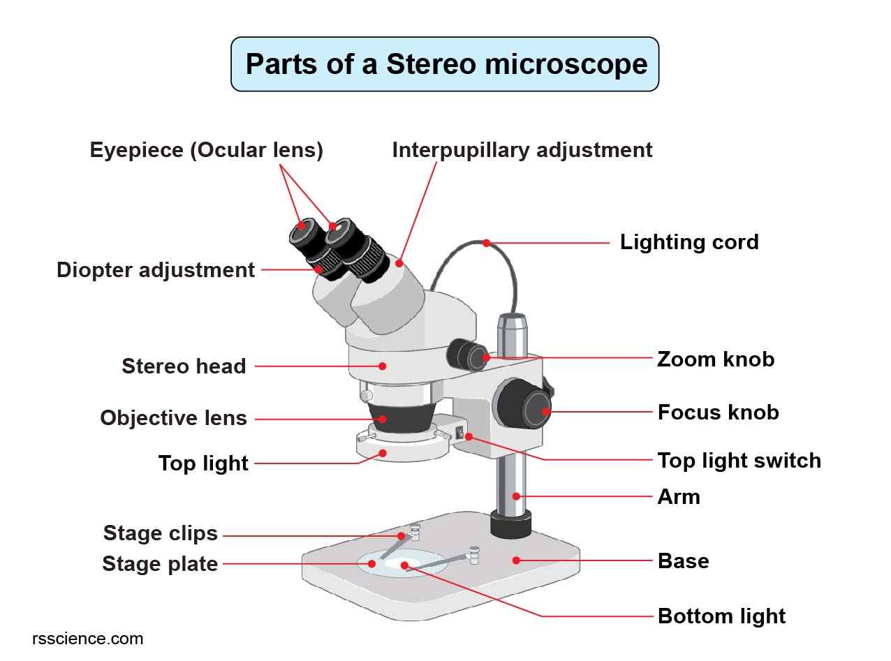
Parts of Stereo Microscope (Dissecting microscope) labeled diagram
The microscope in Figure 1 is illuminated through the oil lamp and water-filled spherical reservoir (also illustrated in Figure 1). Light from the lamp is diffused when it passes through the reservoir and is then focused onto the specimen with a lens attached to the reservoir. This early microscope suffered from chromatic (and spherical.
Cells and Microscopes
Place the slide on the stage and secure it with the stage clip.; Use the coarse focus knob to move the stage as high as it can go. Use stage adjustment knobs to center the "e" so that the light from the light source can pass through it.; Looking through the ocular lenses, lower the stage with the coarse focus adjustment knob until the "e" comes into view.
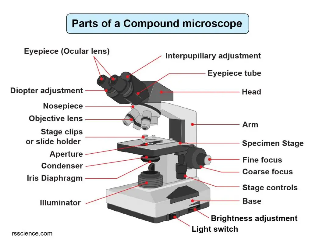
Compound Microscope Parts Labeled Diagram and their Functions Rs
A Study of the Microscope and its Functions With a Labeled Diagram - Science Struck A Study of the Microscope and its Functions With a Labeled Diagram To better understand the structure and function of a microscope, we need to take a look at the labeled microscope diagrams of the compound and electron microscope.
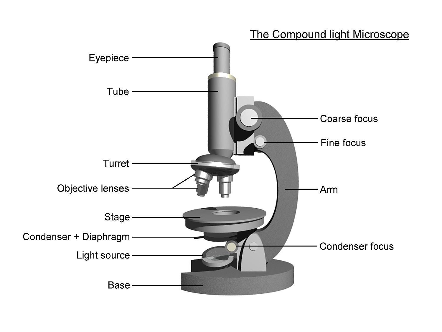
Monday September 25 Parts of a Compound Light Microscope
A labeled diagram of microscope parts furnishes comprehensive information regarding their composition and spatial arrangement within the microscope, enabling researchers to comprehend their function effectively. In this comprehensive article, we will delve into the intricate parts of the microscope, exploring their functions in detail.

301 Moved Permanently
The optical microscope often referred to as the light microscope, is a type of microscope that uses visible light and a system of lenses to magnify images of small subjects. There are two basic types of optical microscopes: Simple microscopes. Compound microscopes. The term "compound" in compound microscopes refers to the microscope having.

Microscope diagram Tom Butler Technical Drawing and Illustration
Magnification is a measure of how much larger a microscope (or set of lenses within a microscope) causes an object to appear. For instance, the light microscopes typically used in high schools and colleges magnify up to about 400 times actual size. So, something that was 1 mm wide in real life would be 400 mm wide in the microscope image.

How to Use a Microscope (Properly) Step by Step New York Microscope
1. Eyepiece 2. Body tube/Head 3. Turret/Nose piece 4. Objective lenses 5. Knobs (fine and coarse) 6. Stage and stage clips 7. Aperture 9. Condenser 10. Condenser focus knob 11. Iris diaphragm 12. Diopter adjustment 13. Arm 14. Specimen/slide 15. Stage control/stage height adjustment 16. On and off switch 17. Base

5 Types of Microscopes with Definitions, Principle, Uses, Labeled Diagrams
Parts of the Microscope (Labeled Diagrams) By Editorial Board December 14, 2022 The microscope is one of the must-have laboratory tools because of its ability to observe minute objects, usually living organisms that cannot be seen by the naked eyes. It is categorized into two: simple and compound microscopes.
The Wonders Of Microscopes What You Need To Know Creyentes Diverses News
Download the Label the Parts of the Microscope PDF printable version here. Download the Label the Parts of the Microscope: Answers PDF printable version here. Microscope World explains the parts of the microscope, including a printable worksheet for schools and home.

Parts of a Microscope The Comprehensive Guide Microscope and
There are three major structural parts of a compound microscope. The head includes the upper part of the microscope, which houses the most critical optical components, and the eyepiece tube of the microscope. The base acts as the foundation of microscopes and houses the illuminator. The arm connects between the base and the head parts.
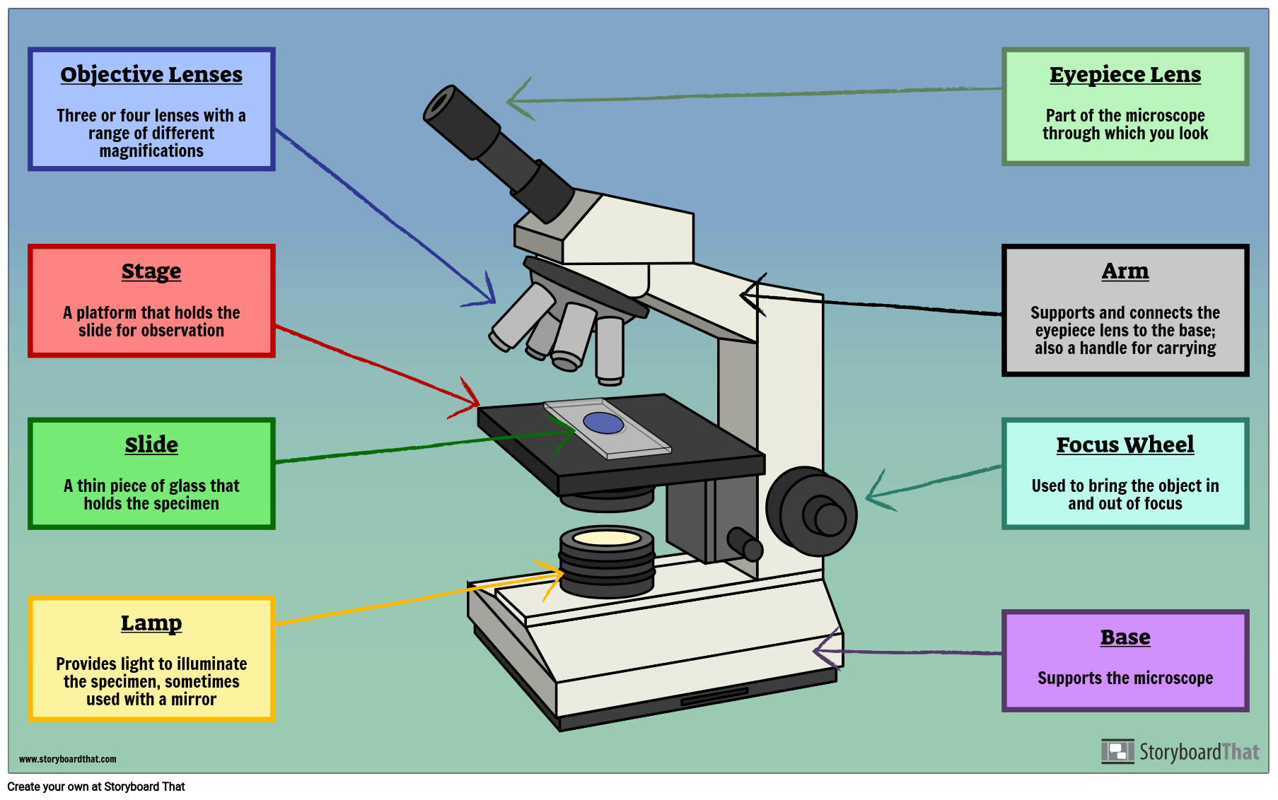
Labelled Microscope with Functions Storyboard by oliversmith
1. Eyepiece or Ocular Lens Eyepiece lens magnifies the image of the specimen. This part is also known as ocular. Most school microscopes have an eyepiece with 10X magnification. 2. Eyepiece Tube or Body Tube The tube hold the eyepiece. 3. Nosepiece Nosepiece holds the objective lenses and is sometimes called a revolving turret.